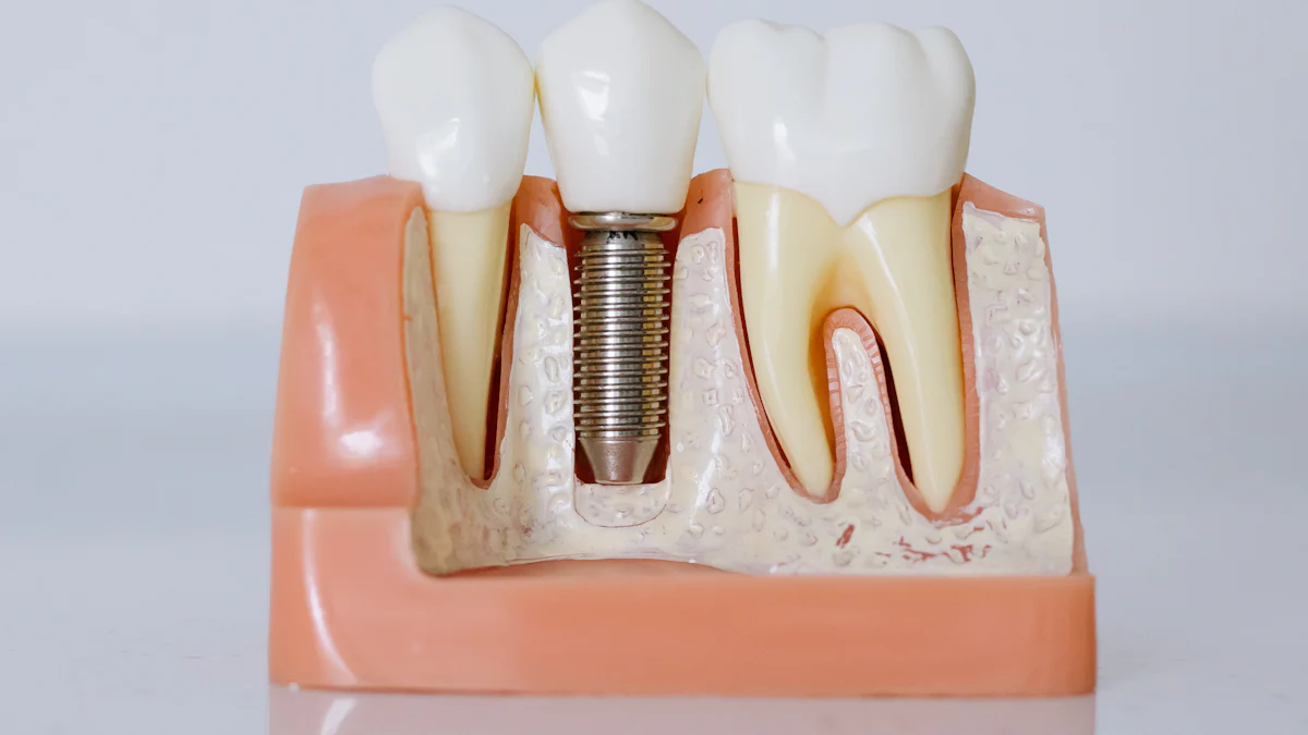
Comparison of Free Gingiva and Attached Gingiva in Dentistry

In dentistry, gingiva refers to the soft tissue that surrounds and protects your teeth. It plays a vital role in maintaining oral health. Free gingiva, also called marginal gingiva, is the portion that lies against the tooth and extends coronally from the free gingival groove. It forms a collar-like band around each tooth but remains unattached to the tooth surface. In contrast, attached gingiva extends apically from the free gingiva to the alveolar mucosa. This portion is firmly bound to the underlying bone and provides structural support.
The primary difference lies in their structure and function. Free gingiva acts as a protective barrier around the teeth, while attached gingiva ensures stability and shields the surrounding tissues. Both types are essential for periodontal health. They prevent gum recession, protect against oral biofilm, and maintain the integrity of the gingival sulcus. Studies demonstrate that a minimum of 2 mm of gingiva is necessary to avoid inflammation, even with good oral hygiene.
Anatomy and Location
Understanding the anatomy and location of the gingiva is essential for appreciating its role in oral health. The gingiva consists of two distinct parts: the free gingiva and the attached gingiva. Each has unique characteristics and functions that contribute to the overall structure of the gingiva.
Free Gingiva
Definition and Characteristics
The free gingiva, also known as marginal gingiva, forms a collar-like band around the neck of each tooth. This portion of the gingiva is not directly attached to the tooth or underlying bone, allowing it to remain flexible. It has an undulating contour and is closely adapted to the tooth surface, creating a rounded margin. The free gingiva is lined by sulcular epithelium, which helps form the gingival sulcus—a shallow space between the tooth and the gum. In healthy individuals, the sulcus depth typically ranges from 1 to 3 millimeters.
Location Around the Teeth
You can observe the free gingiva surrounding the neck of each tooth. It extends coronally from the free gingival groove, a slight depression that demarcates it from the attached gingiva. This groove is not always visible but serves as a distinguishing feature. The free gingiva plays a critical role in forming the gingival sulcus, which is essential for maintaining periodontal health.
The Role of the Free Gingival Groove
The free gingival groove marks the boundary between the free gingiva and the attached gingiva. Although it is not always prominent, this linear depression is an important anatomical landmark. It separates the unattached portion of the gingiva from the firmly bound attached gingiva. The groove also helps clinicians assess the health of the gums during periodontal examinations.
Attached Gingiva
Definition and Characteristics
The attached gingiva lies apically to the free gingiva and is firmly bound to the underlying alveolar bone. This portion of the gingiva is keratinized, making it more resistant to mechanical forces such as chewing and brushing. Its firm attachment provides structural support and stability to the teeth and surrounding tissues. The attached gingiva typically ranges in height from 3 to 12 millimeters, depending on the individual and the location in the mouth.
Location and Boundaries
The attached gingiva extends from the free gingival groove to the mucogingival junction, where it transitions into the alveolar mucosa. This boundary is clearly demarcated and serves as a guide for dental professionals during clinical assessments. The attached gingiva is located adjacent to the free gingiva and plays a vital role in protecting the underlying bone and periodontal ligament.
Variations in Width Across the Mouth
The width of the attached gingiva varies depending on its location in the oral cavity. It is widest in the anterior region, particularly around the incisors, and narrows as it moves toward the posterior teeth. For example, studies demonstrate that the attached gingiva is typically 4 to 5 millimeters wide in the maxillary anterior region but may be as narrow as 2 millimeters in the mandibular premolar area. These variations are normal and do not necessarily indicate any clinical issues.
Function and Role in Oral Health
The gingiva plays a crucial role in maintaining the health and functionality of your oral cavity. Both free gingiva and attached gingiva contribute uniquely to protecting your teeth and surrounding tissues. Understanding their specific functions can help you appreciate their importance in oral health.
Free Gingiva
Role in Protecting the Tooth
The free gingiva acts as the first line of defense for your teeth. It forms a protective collar around the tooth, shielding it from harmful bacteria and debris. This portion of the gingiva creates a seal that prevents food particles and oral biofilm from entering the gingival sulcus. By maintaining this barrier, the free gingiva helps reduce the risk of periodontal disease and gum inflammation.
Contribution to the Gingival Sulcus
The free gingiva plays a vital role in forming the gingival sulcus, a shallow space between the tooth and the gum. This sulcus, lined by sulcular epithelium, allows for the removal of debris during brushing and flossing. A healthy sulcus depth, typically 1 to 3 millimeters, ensures that your gums remain free from infection. The free gingiva's flexibility also allows it to adapt to the tooth's surface, maintaining the integrity of this space.
Attached Gingiva
Role in Providing Structural Support
The attached gingiva provides essential structural support to your teeth and surrounding tissues. Its firm attachment to the alveolar bone ensures stability during chewing and speaking. This portion of the gingiva also:
Acts as a physical barrier to oral biofilm.
Dissipates masticatory forces to protect the underlying bone.
Prevents gingival recession, root exposure, and root caries.
Shields the periodontium from injury caused by external forces.
Importance in Chewing and Stability
The attached gingiva plays a critical role in chewing by absorbing and distributing the forces exerted during mastication. Its keratinized surface resists mechanical stress, ensuring that your gums remain intact even under pressure. This stability allows you to chew comfortably without risking damage to the underlying tissues.
Defense Against External Harm
The attached gingiva employs multiple mechanisms to defend against external harm. These include:
Mechanism Type | Description |
|---|---|
Toughened mechanically resistant surface. | |
Chemical Barrier | Release of antimicrobial substances. |
Biological Barrier | Junctional epithelium forms an epithelial barrier against plaque bacteria. |
Additionally, the attached gingiva protects itself through:
Rapid turnover of cells, which exfoliate harmful bacteria.
Release of cytokines from epithelial cells to combat infections.
Local inflammatory responses that act as a barrier to bacteria and toxins.
These defense mechanisms ensure that your gums remain healthy and resilient, even when exposed to potential irritants.
Clinical Significance
Understanding the clinical significance of free and attached gingiva helps you appreciate their role in dental procedures and oral health. These tissues are not just structural components; they actively contribute to the stability and protection of your teeth and gums.
Importance in Dental Procedures
Role in Periodontal Assessments
Free and attached gingiva play a critical role in periodontal assessments. The free gingiva, closely adapted to the tooth surface, helps form the gingival sulcus. This shallow space, typically 1 to 3 millimeters deep, is a key indicator of gum health. During evaluations, dentists measure sulcus depth to detect early signs of periodontal disease.
The attached gingiva, firmly bound to the alveolar bone, provides a stable foundation for the teeth. A narrow zone of attached gingiva can lead to attachment loss and gingival recession, especially in areas with biofilm accumulation. Studies have observed higher gingival index scores in teeth with a narrow attached gingiva compared to those with a wider zone. Maintaining proper plaque control is essential, but many patients struggle with consistency, increasing the risk of gum disease.
Impact on Restorative and Cosmetic Dentistry
The attached gingiva is vital in restorative and cosmetic dentistry. It acts as a physical barrier to oral biofilm and protects the periodontium from injury. A wide zone of attached gingiva ensures stability during procedures like crown placement or implant surgery. Research demonstrates that implants with less than 2 mm of attached gingiva have higher plaque index scores and are more prone to inflammation.
In cosmetic dentistry, insufficient gingival width can lead to aesthetic concerns like gingival recession and root exposure. A minimum of 2 mm of gingiva (1 mm free and 1 mm attached) is recommended to maintain health and prevent complications. The attached gingiva also dissipates masticatory forces, reducing the risk of root caries and gum damage.
Common Issues and Treatments
Problems Associated with Free Gingiva
Free gingiva is susceptible to inflammation and infection due to its proximity to the gingival sulcus. Poor oral hygiene can lead to plaque buildup, causing gingivitis. This condition results in redness, swelling, and bleeding. If untreated, it may progress to periodontitis, a severe gum disease that damages the supporting structures of your teeth.
Problems Associated with Attached Gingiva
The attached gingiva faces unique challenges. Common issues include:
Gingivitis: Plaque accumulation often triggers inflammation, leading to redness and swelling.
Gingival Recession: Aggressive brushing or periodontal disease can cause gum tissue loss, exposing the tooth's root.
Gingival Hyperplasia: Overgrowth of gum tissue, often linked to medications or poor oral hygiene, can interfere with oral function.
Treatment Options for Gingival Issues
Several treatment options address problems with free and attached gingiva:
Description | |
|---|---|
Free Gingival Grafts (FGGs) | Strengthens soft tissue without significant root coverage, preventing future recession. |
Connective Tissue Grafts (CTGs) | Covers exposed roots and augments gingiva, harvested from the palate. |
Acellular Dermal Matrix (ADM) | Provides root coverage using a graft alternative, reducing the need for donor tissue. |
These procedures, often guided by surgical techniques like guided tissue regeneration (GTR), help restore gum health and prevent further complications. Dentists tailor treatments to your specific needs, ensuring optimal outcomes.
Understanding the differences between free gingiva and attached gingiva helps you appreciate their unique roles in oral health. The free gingiva surrounds the neck of each tooth, forming a collar-like band that protects the gingival sulcus. In contrast, the attached gingiva is firmly bound to the underlying bone, providing stability and shielding the periodontium.
Both types of gingiva contribute to maintaining a healthy oral cavity. They create a physical barrier against biofilm, prevent gingival recession, and protect the gums from injury. The attached gingiva also dissipates chewing forces and ensures structural support. These functions make them essential for periodontal health and dental treatments.
Dentists rely on the health of these tissues during procedures like grafts or guided tissue regeneration. A minimum of 2 mm of gingiva, including both free and attached portions, is crucial for preventing inflammation and maintaining gum stability. By understanding their significance, you can better care for your gums and teeth.
FAQ
The FAQ section addresses common questions about free gingiva and attached gingiva. It provides concise answers to help you understand their roles, anatomy, and clinical significance in oral health.
What is the free gingival groove, and why is it important?
The free gingival groove is a slight depression that separates the free gingiva from the attached gingiva. It serves as a key anatomical landmark for dental professionals during periodontal assessments. This groove helps identify the boundary between the unattached and attached portions of the gingiva.
How does attached gingiva differ from free gingiva?
Attached gingiva is firmly bound to the underlying bone, providing structural support. Free gingiva, on the other hand, is unattached and forms a collar-like band around the tooth. Both play unique roles in protecting your gums and teeth from damage and disease.
Why does the width of attached gingiva vary across the mouth?
The width of attached gingiva depends on its location. It is widest in the anterior region, especially around the incisors, and narrows near the posterior teeth. This variation is normal and does not necessarily indicate any oral health issues.
Can gum disease affect the free gingival groove?
Yes, gum disease can impact the free gingival groove. Inflammation or infection may obscure this groove, making it less visible. Regular dental check-ups help monitor the health of this area and prevent periodontal complications.
What treatments are available for gingival recession?
Treatments for gingival recession include free gingival grafts (FGGs), connective tissue grafts, and guided tissue regeneration (GTR). These procedures restore gum health and prevent further tissue loss. Your dentist will recommend the best option based on your specific needs.
How does attached gingiva protect against gum recession?
Attached gingiva provides a firm barrier that resists mechanical forces like brushing and chewing. Its keratinized surface shields the underlying tissues, reducing the risk of gum recession and root exposure. This stability ensures long-term oral health.
What role does the free gingiva play in maintaining the gingival sulcus?
The free gingiva forms the outer margin of the gingival sulcus, a shallow space between the tooth and gum. This space allows for debris removal during brushing and flossing. A healthy sulcus depth prevents bacterial buildup and gum inflammation.
Why is the free gingival groove not always visible?
The free gingival groove may not be prominent in all individuals. Factors like age, gum health, and anatomical variations influence its visibility. However, its presence remains an important feature for distinguishing between free and attached gingiva.
See Also
Understanding The Various Phases Of Gum Disease Progression
Identifying Signs That Gum Disease May Be Impacting You
User Reviews On ProDentim For Optimal Gum And Tooth Health
