What Is Stippling on Gingiva and Its Role in Oral Health
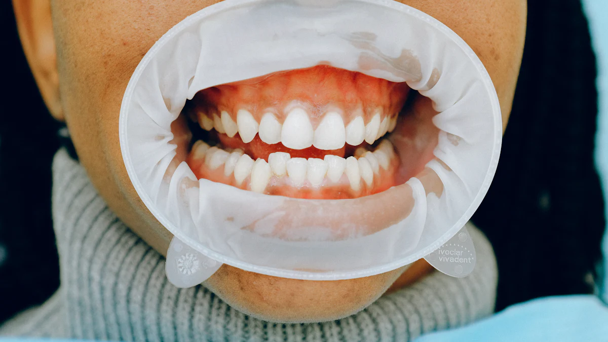
Stippling on gingiva describes the dimpled, orange-peel-like texture observed on the attached gingiva. This distinct surface feature arises from microscopic elevations and depressions formed by connective tissue projections beneath the epithelial layer. Stippling on gingiva is most noticeable on keratinized gingiva, which is tightly adhered to the underlying alveolar bone.
Around 70% of children and 40% of adults display stippling on gingiva, making it a common characteristic of healthy gums. Its presence signifies a well-developed mucosal layer, which enhances gingival stability and resistance to mechanical forces. During oral examinations, clinicians often regard stippling on gingiva as an indicator of normal gingival health.
Understanding Stippling on Gingiva
Stippling on gingiva is a unique feature that reflects the health and structure of the gums. This characteristic texture, often compared to an orange peel, plays a significant role in maintaining gingival stability and overall oral health.
Definition and Appearance
Stippling on gingiva refers to the dimpled or textured surface observed on the attached gingiva. This stippled texture is most prominent on keratinized gingival tissue, which is firmly bound to the underlying alveolar bone. Unlike the freely movable alveolar mucosa, the attached gingiva displays this distinctive pattern due to its structural composition.
The appearance of stippling varies among individuals. It is more pronounced in children and diminishes with age, often disappearing by the age of 50. Stippling is typically visible on the facial surfaces of the gingiva and contributes to its aesthetic appeal. While its presence often indicates healthy gums, smooth gingiva can also be normal if it has always lacked stippling. However, the loss of stippling in previously stippled areas may signal inflammation or disease, such as gingivitis.
Anatomical Basis
Role of Connective Tissue and Epithelial Layers
The stippled texture arises from the interaction between the connective tissue and epithelial layers of the gingiva. Microscopic elevations and depressions on the gingival surface create this pattern. These features result from connective tissue projections, known as papillae, which align with epithelial ridges called rete pegs. This structural arrangement strengthens the gingiva and enhances its resistance to mechanical forces.
The connective tissue contains collagen fibers that anchor the gingiva to the underlying bone and cementum. This firm attachment ensures stability and prevents the gingiva from shifting during chewing or brushing. The stippling process reflects the intricate relationship between these layers, highlighting the complexity of periodontal anatomy.
Connection to Keratinized Gingiva
Stippling is closely associated with keratinized gingival tissue. Keratinization refers to the presence of a tough, protective layer on the gingival surface. Higher levels of keratinization correlate with more pronounced stippling. This keratinized tissue provides a durable barrier against bacterial invasion and mechanical stress, contributing to the overall health of the periodontium.
The attached gingiva, where stippling occurs, plays a critical role in oral health. Its firm connection to the alveolar bone and keratinized surface ensures that the gingiva remains resilient and functional. This unique texture not only supports the gums but also serves as a visual indicator of their health.
Stippling on Gingiva and Oral Health
Stippling on gingiva serves as more than just a visual feature; it plays a vital role in maintaining oral health. Its presence reflects the structural integrity of the gingival tissues and their ability to resist external challenges.
Indicator of Healthy Gums
Association with Firm, Attached Gingiva
The stippled texture of gingiva often indicates firm, attached gingiva. This type of gingiva is tightly bound to the underlying alveolar bone and cementum, ensuring stability during daily activities like chewing and brushing. The firm attachment prevents the gingiva from shifting, which helps maintain the natural contour of the gums. Clinicians frequently evaluate stippling during oral examinations as a sign of healthy gingival tissues.
Resistance to Mechanical Forces and Bacterial Invasion
Stippling contributes to the gingiva's ability to withstand mechanical forces. The microscopic elevations and depressions create a robust surface that resists trauma from brushing or mastication. Additionally, the keratinized layer associated with stippling acts as a barrier against bacterial invasion. This protective feature reduces the risk of infections, such as gingivitis, and supports overall gingival health.
Protective Role
Contribution to Gingival Stability
The stippled appearance of gingiva enhances its stability. The connective tissue projections, or papillae, interlock with epithelial ridges to create a firm bond. This structural arrangement minimizes the risk of gingival detachment, which could expose the underlying bone and roots to harmful elements. The stability provided by stippling ensures that the gingiva remains functional and resilient over time.
Role in Maintaining Periodontal Health
Gingival stippling plays a crucial role in preserving periodontal health. Studies have shown that areas with stippling often exhibit greater keratinized tissue width (KTW), buccal bone thickness (BT), and gingival thickness (GT). These parameters contribute to a stronger periodontium, which protects the teeth and surrounding structures.
Finding | Description |
|---|---|
Correlation with KTW | Statistically significant association between stippling and keratinized tissue width. |
Correlation with BT | Greater buccal bone thickness observed in areas with stippling. |
Correlation with GT | Increased facial gingival thickness in regions exhibiting stippling. |
Prevalence of Stippling | 56% of men and 44% of women showed facial stippling; more common in central incisors. |
Interdental Stippling Prevalence | 73% of interdental sites exhibited stippling, particularly in men and central incisor regions. |
The protective role of stippling extends to interdental areas, where it frequently appears. These regions benefit from the added stability and resistance provided by stippled gingiva, reducing the likelihood of periodontal disease.
Gingival stippling varies among individuals in size, number, and distribution. While its presence often indicates health, its absence does not always signify disease.
Factors Influencing Stippling on Gingiva
Several factors influence the presence and characteristics of stippling on gingiva. These include age, oral hygiene, inflammation, and genetic variability. Understanding these factors helps clinicians evaluate gingival health and identify potential concerns.
Age and Development
Prominence in Children Versus Adults
Stippling on gingiva first appears around the age of six. It becomes more prominent during adolescence when the gingival tissues are at their healthiest. Children often exhibit a more pronounced stippled texture due to the firmness and resilience of their attached gingiva. This characteristic diminishes as individuals grow older.
Reduction with Aging
As people age, the stippled appearance of the gingiva tends to fade. This reduction occurs due to changes in the connective tissue and epithelial layers. The gingival texture becomes smoother, and the attached gingiva may lose some of its firmness. Studies show that stippling is less common in adults over 50, reflecting the natural aging process and its impact on gingival structure.
Oral Hygiene and Inflammation
Impact of Plaque and Gingivitis
Poor oral hygiene can lead to plaque buildup, which causes gingivitis. Infected or inflamed gingiva often loses its stippled texture. The smooth, swollen appearance of the gums during inflammation contrasts with the normal dimpled surface. This change serves as an early warning sign of gingival disease.
Restoration with Improved Oral Care
Improved oral hygiene can restore the stippled appearance of gingiva in some cases. Regular brushing, flossing, and professional cleanings help reduce inflammation and promote healthy gingival tissues. The return of stippling indicates the resolution of gingivitis and the restoration of gingival health.
Genetic and Individual Variability
Differences Among Individuals
The presence and prominence of stippling vary significantly among individuals. Males generally exhibit more pronounced stippling than females. The upper labial gingiva often shows the heaviest stippling, while other areas may display lighter or no stippling. These differences highlight the role of individual anatomical characteristics in determining stippling patterns.
Influence of Genetic Predisposition
Genetic factors play a crucial role in the presence of stippling. The width of attached gingiva, root prominences, and other anatomical features influence the stippled texture. These traits are often inherited, making stippling a unique characteristic for each person. Research shows that genetic predisposition affects the size, number, and distribution of stippling, emphasizing its individualized nature.
The presence of stippling on gingiva reflects a combination of developmental, environmental, and genetic factors. While its absence does not always indicate disease, changes in stippling can signal underlying issues that require attention.
Clinical Significance of Stippling on Gingiva
The clinical significance of stippling on gingiva extends beyond its aesthetic appeal. It serves as a valuable diagnostic tool and provides insights into the health of gingival tissues. However, its variability among individuals highlights the need for a comprehensive approach when assessing gingival health.
Diagnostic Value
Use in Assessing Gingival Health
The presence of stippling offers a reliable indicator of gingival health. Clinicians often evaluate stippling during routine oral examinations to determine the firmness and attachment of gingival tissues. Various diagnostic methods have been employed to study stippling and its characteristics.
Diagnostic Method | Source |
|---|---|
Clinical Photographic Evaluation | Greene studied the stippling through clinical photographic and histologic evaluation. |
Histologic Evaluation | Greene studied the stippling through clinical photographic and histologic evaluation. |
Casts and Impressions | Rosenberg additionally utilized the casts and impressions to study the stippling. |
Negative Replica Method | Vendrine and others utilized a negative replica method to study the stippling. |
These methods help clinicians assess the texture, distribution, and prominence of stippling, which can reflect the underlying health of the gingiva.
Absence as a Potential Sign of Disease
The absence of stippling may indicate gingival inflammation or other conditions. Infected or inflamed gingiva often loses its stippled texture, appearing smooth and swollen. However, smooth gingiva does not always signify disease unless it results from the loss of previously existing stippling. Once gingival health is restored, stippling can reappear, signaling the resolution of inflammation.
The absence of stippling on gingiva can occur due to gingivitis or other inflammatory conditions. Its reappearance often indicates improved oral health.
Variability and Limitations
Not Always Present in Healthy Individuals
Stippling is not universally present, even in healthy individuals. Factors such as age, sex, and genetic predisposition influence its appearance. Males tend to exhibit more pronounced stippling compared to females. Additionally, stippling diminishes with age, becoming less visible in older adults. These variations limit its reliability as a sole indicator of gingival health.
Importance of Considering Other Clinical Factors
Clinicians must consider additional factors when evaluating gingival health. The width of attached gingiva, root prominences, and keratinization levels all contribute to the presence of stippling. Variations in stippling characteristics can provide insights into an individual’s gingival health. However, small sample sizes in studies and individual differences highlight the need for a holistic approach.
Gingival stippling is influenced by the width of attached gingiva.
Root prominences affect stippling characteristics.
Age and sex differences impact the distribution and appearance of stippling.
Males often display heavier stippling compared to females.
Stippling on gingiva remains a valuable diagnostic feature, but its variability underscores the importance of considering other clinical parameters during evaluations.
Stippling on gingiva, characterized by its orange-peel-like texture, reflects the intricate relationship between connective tissue and epithelial layers. This unique feature, primarily found on attached gingiva, serves as a marker of healthy gums. Its presence indicates firm, keratinized tissue that resists bacterial invasion and mechanical forces.
Key Anatomical Characteristics of Stippling
Characteristic | Description |
|---|---|
Texture | Resembles an orange peel, indicative of healthy gingiva. |
Location | Present on the attached gingiva, absent on the alveolar mucosa. |
Variation | Varies in number, size, and prominence among individuals based on age, sex, and gingival width. |
Diagnostic Significance | Presence indicates a well-developed mucosal layer; loss suggests gingival inflammation. |
Surface Comparison | More prominent on the facial surface than the lingual side. |
Stippling contributes to oral health by enhancing gingival stability and protecting underlying structures. Its association with keratinization supports the periodontium and aids in early diagnosis of conditions like gingivitis. Factors such as age, sex, and gingival width influence its presence, with males and younger individuals often displaying more pronounced stippling.
The presence or absence of stippling offers valuable insights into gingival health. Clinicians should consider its variability and evaluate other parameters during examinations.
FAQ
What causes stippling on gingiva?
Stippling on gingiva occurs due to microscopic elevations and depressions formed by connective tissue papillae and epithelial ridges. This structural arrangement strengthens the gingiva, creating its characteristic orange-peel texture. Factors like keratinization and the width of attached gingiva also contribute to its appearance.
Is stippling on gums a sign of healthy gingiva?
Yes, stippling on gums often indicates healthy gingiva. It reflects firm, keratinized tissue that resists bacterial invasion and mechanical forces. However, its absence does not always mean disease unless it results from the loss of previously existing stippling due to conditions like gingivitis.
Why does stippling diminish with age?
Stippling diminishes with age due to changes in the connective tissue and epithelial layers. The gingiva becomes smoother and less firm over time. This natural process often leads to a reduction in the stippled texture, especially in adults over 50.
Can poor oral hygiene affect gingival stippling?
Yes, poor oral hygiene can lead to plaque buildup and gingivitis, causing the loss of gingival stippling. Inflamed gums appear smooth and swollen instead of stippled. Improved oral care, including brushing and flossing, can help restore the stippled texture in some cases.
Does everyone have stippling on their gingiva?
No, not everyone has stippling on their gingiva. Its presence varies among individuals based on factors like age, sex, and genetic predisposition. While males and younger individuals often display more pronounced stippling, its absence can still be considered normal in some cases.
Stippling on gingiva serves as a valuable indicator of gum health but varies widely among individuals. Regular dental check-ups ensure proper evaluation of gingival health.
See Also
The Link Between Stress, Gum Disease, And Oral Wellness
Exploring The Various Phases Of Gum Disease
The Effects Of Pro Dentin Candy On Dental Health